Free Republic
Browse · Search
News/Activism
Topics · Post Article

To: Westlander
Don’t have to. After 30+ years as a registered CT tech in Level 1 trauma centers in an urban environment on the midnight shift, I can ‘read’ the crap out of CT’s-neuro, body, and ortho. By the way, one of the ‘comparative’ images is actually a T1 MRI sagital reconstruction-not a CT.

Are you saying that Terri Schiavo had more or less liquid in her brain vs. working brain tissue?

75 posted on 07/20/2007 4:44:09 PM PDT by Spiff (Rudy Giuliani Quote (NY Post, 1996) "Most of Clinton's policies are very similar to most of mine.")
[ Post Reply | Private Reply | To 69 | View Replies ]


To: Spiff
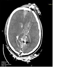
Hydrocephalus is not the lone deciding factor for ascertaining brain viability. Image below is an example. Note no enlarged ventricles, although they have fresh blood shown on this one slice. Entrance wound right occiput, bone and metal fragments thruout both lobes, exit wound thru left frontal bone. (images are interpeted as if the patient is facing you) This type of penetrating wound are usually assigned to second year neurosurgery residents for 'practice' in the O.R. Survivability is zero. Yet, as shown, there is no hydrocephalus. It takes a team--neurosurgery, neurology, diagnostic radiology, to perform a battery of exams to find a diagnosis and impending outcome for the patient.
92 posted on 07/20/2007 6:29:38 PM PDT by Westlander (Unleash the Neutron Bomb)
[ Post Reply | Private Reply | To 75 | View Replies ]

Free Republic
Browse · Search
News/Activism
Topics · Post Article


FreeRepublic, LLC, PO BOX 9771, FRESNO, CA 93794
FreeRepublic.com is powered by software copyright 2000-2008 John Robinson