Free Republic
Browse · Search
Smoky Backroom
Topics · Post Article

To: Peach; Howlin; onyx; All
On Greta's show last night, she asked a brain specialist to explain the cat scan of Terri's brain.

They had a brain scan of a healthy brain next to Terri's.

This doctor said Terri's brain is 80% liquefied. The 20% remainder is damaged. Nothing viable, nothing there.

(I tried to see if Greta had a transcript but she doesn't, because I would like to have the exact words this Doctor Pecotpht[sp?] used.

Anyway, what he said is what I figured was the case days ago.

Just looking at the two brain comparisons said it all.

Other than a brain transplant or growing a new one, Terri's brain is not viable. Any "thearpy" would be beyond futile. Waste of time.

Even a new scan, PET done today would only show the brain to be further deteriorated. No way has it "improved" since the last scan.

972 posted on 03/25/2005 9:44:51 AM PST by the Deejay ( I'LL RESPECT YOUR OPINION....IF YOU'LL RESPECT MINE.....)
[ Post Reply | Private Reply | To 947 | View Replies ]


To: the Deejay; daylate-dollarshort

The destruction of Terri's brain is worse than has been previously described. The Neurologist that was interviewed stated that 80% of Terri's brain has been destroyed and has been replaced by fluid. He further stated that there is no way that she is not in PVS and further, it is impossible that she will ever improve.

I appologize for the quality of the pic, but I was lucky to get this.

528 posted on 03/24/2005 11:12:53 PM EST by daylate-dollarshort (s/v Musashi I)

976 posted on 03/25/2005 9:48:26 AM PST by Howlin
[ Post Reply | Private Reply | To 972 | View Replies ]

To: the Deejay; Howlin


See #976 -- I've seen the two side by side.
Howlin has it posted.


980 posted on 03/25/2005 9:51:20 AM PST by onyx (Robert Frost "Good fences make good neighbors." Build the fence, Mr. President and Congress.)
[ Post Reply | Private Reply | To 972 | View Replies ]

To: the Deejay

FOX News, March 24, 2005

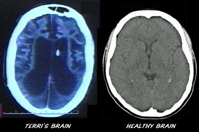
Experts: CT shows little gray matter


By Bob Lamendola

Fort Lauderdale, Fla. — Scans of Terri Schiavo's brain show that the great majority of her gray matter where thinking and feeling occur has died off and been replaced by watery fluid, with no chance of growing back, neurologists said.

About 70 percent to 90 percent of Schiavo's upper brain is gone, and there's also damage to her lower brain that controls instinctive functions like breathing and swallowing, said three Florida neurologists who viewed 12 of her CT "computed tomography" X-ray scans.

"This is as severe brain damage as I've ever seen," said Dr. Leon Prockop, a professor and former chairman of neurology at the University of South Florida College of Medicine in Tampa, upon viewing the scans.

Dr. Walter Bradley, chairman of neurology at the University of Miami's Miller School of Medicine, added: "I doubt there's any activity going on in the higher levels of her brain."

The assessments came as a neurologist for the state who saw the CT scans and examined Schiavo said he saw signs of awareness in her behavior and concluded that more extensive tests should be done.

Dr. William Chesire Jr. made his assertions in an affidavit that Gov. Jeb Bush used Wednesday to ask a court to reinsert her feeding tube while the state investigates. Cheshire sees patients at the Mayo Clinic in Jacksonville and teaches at Trinity International University, a Christian institution. It's the latest example of specialists disagreeing over Schiavo's condition.

The CT scans the doctors examined were filed in court as part of the protracted legal fight over whether to disconnect Schiavo's feeding tube.

The Sun-Sentinel asked the three neurologists to view the scans via e-mail. They have not examined Schiavo personally and had no role in the court case. The scans were taken in 1996, five years after her heart stopped due to a chemical imbalance cutting off blood to her brain and leaving her severely incapacitated for the past 15 years.

The three neurologists said a scan of a normal upper brain, or cerebral cortex, is almost totally gray. Schiavo's scans show large black patches, which the doctors said were areas where cells have died due to a lack of oxygen and the tissue has shrunk. The dead cells get broken down and taken away, and those areas have filled with pools of cerebrospinal fluid, the doctors said.

The neurologists said they can't tell from a CT scan whether remaining tissue is functioning, but all three said they doubted that the neurons — brain cells responsible for thought — could survive the loss of oxygen that Schiavo sustained.


996 posted on 03/25/2005 10:01:30 AM PST by daylate-dollarshort (s/v Musashi I)
[ Post Reply | Private Reply | To 972 | View Replies ]

To: the Deejay; Howlin
Comparison of Terri Schiavo's brain to a healthy brain.

The area shown in black in this diagram is the area damaged in Terri Schiavo.

About the only portion of her brain that is still mostly intact is the brain stem -- that portion which connects to the spinal cord and which process automatic functions such as breathing, blinking, eye movement. It is the area shown in black in this image.

Anyone who has been claiming that she can speak, respond to commands, acknowledge the presence of others, etc., is either lying or deluding themselves. This next photo is a cutaway view of a human brain in a cadaver. This cadaver's brain looks more intact and healthier than Terri Schiavo's brain.

In her current state, Terri Schiavo is not too much different from an anencephalic baby (a baby born without most or all of its brain).

1,051 posted on 03/25/2005 10:26:10 AM PST by Wolfstar (If you can lead, do it. If you can't, follow. If you can't do either, become a Democrat.)
[ Post Reply | Private Reply | To 972 | View Replies ]

To: the Deejay
Toskrin is a neurologist and member of Free Republic

At: Feeding Tube Removed: Greer Gives Order - Toskrin's reply no. 2021:

I'm a neurologist who has followed this case from time to time; it's been about a year since I looked into it though.

The neurologic question seems to be if she if a persistent vegetative state (PVS) or what is called a minimally conscious state (MCS). From the videos I've seen, she probably is MCS, but just barely. In her case it's hard to be sure without actually being there during the exam. However, I understand that the videos are a few years old and that her condition may have deteriorated since.

I believe she has been examined by court-appointed neurologists who claim that she is PVS. That may be the case if she is now worse than the videos suggest, or if the videos are misleading. Unfortunately, her parents have chosen to associate with a group of doctors who are, to be polite, out of the mainstream in their practices and I don't trust their evaluations.

The bigger issue to me is whether Michael is competent to act in her best interests. Much of what he has done in the intervening time suggests that he is not.

I think if anyone has dropped the ball here, it is the Florida legislature and governor. They have had plenty of time to pass laws addressing the issue of next-of-kin and what to do when a spouse or child may not be acting in the best interests of the patient.

At: Feeding Tube Removed: Greer Gives Order - Toskrin's reply to supercat, no. 2053:

What should be clear, though, is that a 45-minute examination by a doctor is not enough to make a diagnosis that someone is in an incurable vegetative state.

Agreed, but the length of the exam is not the most important thing. The patient will probably be the same at the start of the forty-five minutes as at the end. More important is seeing the patient over time, on different days and at different times. The doctor also has to rely on the observations of reliable staff for the assessment. If five nurses tell you the patient talked, then they're not PVS, even if the doctor didn't see it.

At: Doctor: The "Water" in Terri’s Brain is a Myth [CT Scan Found As Well As Bone Scan Of Terri] - Toskrin's reply no. 31:

I'm a neurologist and have read hundreds of CT scans. Two problems with this scan are that there is no name on it and it is only a single image of the multiple image complete CT scan.

That being said, I don't see any cortex on this CT image. There is white matter but the normal "cortical ribbon" that surrounds the white matter is missing.

At: Another Doctor in Support of Terri - Toskrin's reply no. 74:

Finally, we have the report of a disinterested neurologist whos affidavit is in line with what is observed on the videotapes and gathered from other testimony. His summary is a million times more credible than either Hammesfahr's or Cranford's.

Here's a link to his bio:

http://www.mayoclinic.org/neurology-jax/11736755.html

At: Neurologists see little sign of activity in Schiavo's brain - Toskrin's reply no. 251:

[A] conventional brain MRI is not definitive at all and would add little to the argument one way or another. A PVS diagnosis is based primarily upon serial exams over time and is supplemented by functional studies - EEG, PET, and functional MRI. Any functional test, however, is going to be secondary to the results of the examinations.

At: Neurologists see little sign of activity in Schiavo's brain - Toskrin's reply to cajungirl, no. 317:

[A] PET would show areas lighting up in response to stimuli. It shows in living color the function or working of the brain...

Yes, I know. I was just interested in what msuMD * thought was wrong with the statements about PET scanning in the affidavit. I didn't see anything wrong.

I'm a neurologist and have seen thousands of CT scans and I have to say I've also seen lots of elderly people walking and talking who have that amount of brain volume. But that's a little misleading. In Terri's scan, the occipital lobes and parts of the parietal lobes are totally destroyed with no recognizable structure. I can't say I've ever seen anyone walking around like that.

In her case, any structural imaging is irrelevant. If you don't see cortex, that doesn't mean it's not there. If you do, that doesn't mean it's functioning. The recent affidavit is the strongest argument that she is in a minimally conscious state rather than a PVS.

*msuMD's statement, in reply no. 248, had been:

This doctor must re-learn what in fact a PET scan does. First of all a PET uses tagged glucose to measure uptake in the brain. A MRI evaluates structures in the brain....all of which can remain normal in a person post hypoxic encephalopathy....and mean absolutely nothing.

At: Neurologists see little sign of activity in Schiavo's brain - Toskrin's reply no. 329:

I only saw the one picture, not all the images. Yes, you can do a PET with a stimulator. It looks like she has a shunt on the CT, but it's well documented that she also has a thalamic stimulator. She might have both. For what it's worth I would guess that she almost certainly would have had a MRI done in 1990 before the stimulator was placed.

The image:


1,064 posted on 03/25/2005 10:30:57 AM PST by First_Salute (May God save our democratic-republican government, from a government by judiciary.)
[ Post Reply | Private Reply | To 972 | View Replies ]

Free Republic
Browse · Search
Smoky Backroom
Topics · Post Article


FreeRepublic, LLC, PO BOX 9771, FRESNO, CA 93794
FreeRepublic.com is powered by software copyright 2000-2008 John Robinson