Posted on 01/04/2021 1:59:04 PM PST by BenLurkin
Fluorescence microscopy is widely used in biochemistry and life sciences because it allows scientists to directly observe cells and certain compounds in and around them. Fluorescent molecules absorb light within a specific wavelength range and then re-emit it at the longer wavelength range. However, the major limitation of conventional fluorescence microscopy techniques is that the results are very difficult to evaluate quantitatively; fluorescence intensity is significantly affected by both experimental conditions and the concentration of the fluorescent substance. Now, a new study by scientists from Japan is set to revolutionize the field of fluorescence lifetime microscopy.
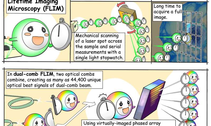
A way around the conventional problem is to focus on fluorescence lifetime instead of intensity. When a fluorescent substance is irradiated with a short burst of light, the resulting fluorescence does not disappear immediately but actually "decays" over time in a way that is specific to that substance. The fluorescence lifetime microscopy technique leverages this phenomenon, which is independent of experimental conditions, to quantify fluorescent molecules and changes in their environment. However, fluorescence decay is extremely fast, and ordinary cameras cannot capture it. While a single-point photodetector can be used instead, it has to be scanned throughout the sample's area to be able to reconstruct a complete 2-D picture from each measured point. This process involves movement of mechanical pieces, which greatly limits the speed of image capture.
In this recent study, published in Science Advances, the team of scientists developed a novel approach to acquire fluorescence lifetime images without the need for mechanical scanning. Professor Takeshi Yasui, from Institute of Post-LED Photonics (pLED), Tokushima University, Japan, who led the study, says, "Our method can be interpreted as simultaneously mapping 44,400 light-based 'stopwatches' over a 2-D space to measure fluorescence lifetimes—all in a single shot and without scanning."
(Excerpt) Read more at phys.org ...
so a phased array receiver for light.... I’m sure there are additional applications for that....
This is a big deal. Good for the Japanese. They are learning to innovate
Disclaimer: Opinions posted on Free Republic are those of the individual posters and do not necessarily represent the opinion of Free Republic or its management. All materials posted herein are protected by copyright law and the exemption for fair use of copyrighted works.